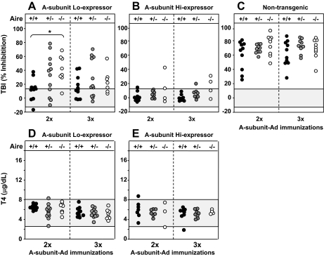
Figure 3.
TSHR antibodies and serum T4 in transgenic mice expressing the human A subunit at low levels (Lo-expressors, A and D) or high levels (Hi- expressors, B and E) or in nontransgenics (C) of the following Aire genotypes: +/+, +/− and −/−. Mice were immunized three times with A-subunit Ad; blood was drawn 1 wk after two immunizations and 4 wk after the third injection. Data are shown for individual mice. A–C, TSHR antibodies measured by TBI (%) in undiluted sera (see Materials and Methods) in Lo and Hi A-subunit transgenics (A and B, respectively) and nontransgenic mice (C). The shaded area represents the mean ± 2 sd values for sera from nontransgenic mice immunized with Con-Ad. Values in Aire −/− Lo-expressor transgenic mice were significantly greater than in Aire +/+Lo expressor transgenics: *, P = 0.002 (t test). Note that because of the near-maximal TBI values for undiluted sera in nontransgenic mice (C), the difference between TBI levels for Aire −/− and +/+ mice after two immunizations was not significantly different. D and E, Serum T4 (μg/dl) in Lo (D) and Hi (E) expressors transgenic for the A subunit of the following Aire genotypes: Aire −/−, Aire +/−, and +/+. T4 values for nontransgenic mice are shown in Fig. 1, D and E. The shaded area represents the mean ± 2 sem values in nontransgenic mice immunized with Con-Ad.

