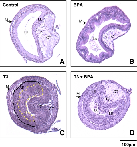
Figure 2.
In the presence of BPA, intestinal remodeling is delayed during T3-induced metamorphosis as early as 4 d after treatment. Tadpoles of the same size and at the same stage (stage 54) were treated with T3 to initiate the metamorphic process. Four days later, the intestines were isolated, fixed, and the sections stained with hematoxylin-eosin. Representative control (A), BPA- (B), T3- (C), and T3+BPA (D)-treated tadpoles are shown. Note that the control, BPA, and T3+BPA intestine remained largely typical of tadpole intestine, as seen by the presence of a thin muscle layer, little connective tissue, and little or no adult intestinal precursor cells. Histology of T3-treated intestines revealed increased muscle layer thickness, proliferation of connective tissue, and the appearance of adult epithelial cells (the larval epithelial cells are surround by a yellow dashed ring, whereas the appearance of adult epithelial cells are represented between a black solid and the yellow dashed line). This experiment was repeated four times with similar results. Scale bar, 100 μm. AE, Adult epithelium; CT, connective tissue; LE, larval epithelium; Lu, lumen; M, muscle; Ty, typhlosole.

