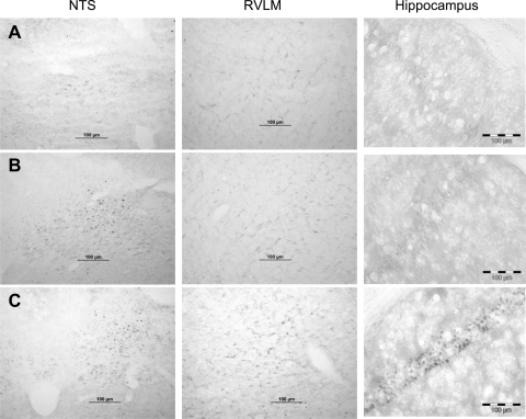
Fig. 1.
Immunohistochemical localization of occupied glucocorticoid receptors (GR). Rats were treated with dorsal hindbrain (DHB) sham (row A) or DHB mifepristone (Mif) (row B) pellets for a total of 6 wk and were adrenalectomized at least 72 h prior to collection of the brains. Each row contains data from a single animal. The rat in row C was adrenal-intact and subjected to restraint stress for 60 min just prior to collection of the brain. Immunohistochemistry was performed using an antibody that has higher affinity for the occupied, compared with the unoccupied, GR. Scale bars are all equal to 100 μm. NTS, nucleus of the solitary tract; RVLM, rostral ventrolateral medulla.

