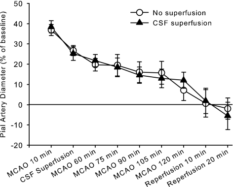
Fig. 1.
Percent change in pial arteriolar diameter (±SE) during 2 h of middle cerebral artery occlusion (MCAO) and 20 min of reperfusion with either no superfusion of the cranial window (n = 9) or continuous superfusion of cerebrospinal fluid (CSF; n = 9). Two-way ANOVA indicated no significant interaction between group treatment and time (P = 0.99).

