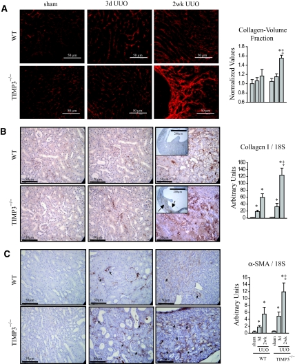
Figure 3.
Increased interstitial fibrosis, enhanced collagen synthesis, and α-smooth muscle actin (αSMA) expression in response to tubulointerstitial injury in tissue inhibitor of matrix metalloproteinases 3 (TIMP3)−/− mice. (A) Picrosirius red staining and confocal microscopy shows marked thickening and disorganization of the peritubular fibrosis in TIMP3-deficient kidneys compared with wild-type (WT) kidneys at 2 wk after unilateral ureteral obstruction (UUO). (B) Enhanced collagen synthesis and deposition as shown by immunostaining for collagen type I (arrows indicate areas of collagen deposition), as well as TaqMan RNA analysis for collagen type I normalized to 18S. (C) αSMA shows a significantly higher degree of staining and mRNA expression in TIMP3−/− kidneys at 3 d and 2 wk post-UUO. Arrows indicate regions of interstitial staining for αSMA. *P < 0.05 compared with sham-operated group; ‡P < 0.05 compared with corresponding WT group.

