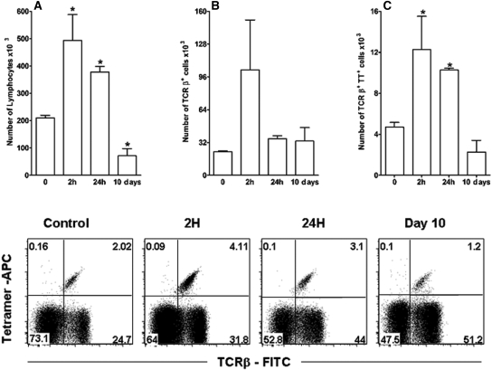
Figure 1.
The development of anti-GBM GN is associated with local influx of iNKT cells. C57BL/6 mice were injected with anti-GBM serum on day 0. At 0, 2, and 24 h and on day 10, kidneys were collected, treated with collagenase and DNase, and stained for TCR-β antigens (TCRβ-FITC) and Vα14-Jα18 TCR (TT, tetramer-CD1d+αGalCer-APC) to detect iNKT cells at different time points. FACS analysis showed the time-dependent influx of lymphocytes (A), TCRβ+ cells (B), and iNKT lymphocytes (C). Results are reported as absolute numbers of infiltrating cells per kidney (mean ± SEM) from five to eight animals for each time point. *P < 0.05 versus 0 h. Data are representative of three separate experiments.

