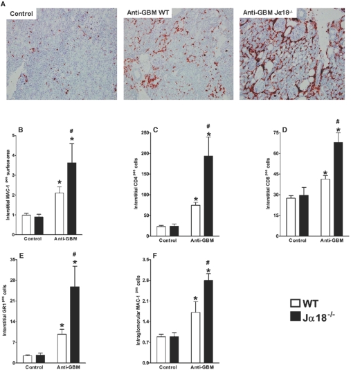
Figure 3.
Increased infiltration of inflammatory cells into renal tissue in mice lacking iNKT cells. C57BL/6 WT and Jα18−/− mice were injected with anti-GBM serum, and control mice were injected with PBS. After 14 d, kidneys were collected. (A) Immunohistochemical analysis showed greater influx of inflammatory cells into renal tissue of Jα18−/− mice, which lack iNKT cells, than into renal tissue from WT mice. MAC-1+ cells (A, B, and F) made up the largest proportion of infiltrating cells, followed by CD4+ T cells (C), CD8+ T cells (D), and GR1+ cells (E). Jα18−/− mice also had more intraglomerular MAC-1+ cells than WT mice (F). Results are expressed as mean ± SEM (n = 5). *P < 0.05 versus control; #P < 0.05 versus WT. This is representative of three isolated experiments.

