Figure 1.
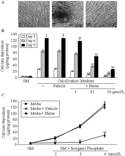
Heme inhibits HSMC calcification induced by elevated Pi in a dosage-dependent manner. (A) HSMCs were cultured in GM (I) or in calcification medium in the absence (II) or presence of heme (50 μmol/L; III) for 9 d. Von Kossa staining of cells was performed as described in the Concise Methods section. Representative picture of three separate experiments. (B) HSMCs were cultured in GM or in calcification medium alone or supplemented with NaOH (Vehicle, 1 mmol/L) or 5, 25, and 50 μmol/L heme (dissolved in NaOH at a final concentration of 1 mmol/L in each group). Calcium contents of cells were measured after 3 (□), 6 (  ), and 9 d (▪) of culture as described in the Concise Methods section and were normalized by protein content. Data are means ± SD of three independent experiments performed in duplicate. (C) HSMCs were cultured in GM alone or supplemented with 2, 3, or 4 mmol/L Pi (•). The media containing different amounts of Pi was supplemented with heme (50 μmol/L; ▴) or with NaOH (vehicle, 1 mmol/L; ▵). Calcium deposition was measured at day 9, and results were normalized by protein content of the cells. Data show the average of three separate experiments performed in duplicate. *P < 0.05; **P < 0.01. Magnification, ×100.

