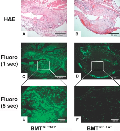
Figure 5.
Bone marrow contribution to the NH lesion was examined by lethal total body irradiation of GFP and wild-type mice followed by GFP bone marrow reconstitution (BMTWT→GFP versus BMTGFP→WT) and renal ablation. (A and B) Six weeks after bone marrow reconstitution, renal ablation was performed, and an AV fistula was created. Three weeks later, venous anastomoses were sectioned and stained for H&E (scale bar, 0.02 mm). (C) Fluorescence detected at 1 s of BMTWT→GFP demonstrated that GFP is densely present in the neointima (scale bar, 0.02 mm). (D) However, fluorescence detected at 1 s of BMTGFP→WT demonstrated that GFP is not detected in the NH lesion and only in the adventitia, suggesting that bone marrow-derived cells do not contribution to the make-up of the NH lesions (scale bar, 0.2 mm). (E and F) Higher magnification with 5 s of fluorescence (scale bar, 0.05 mm).

