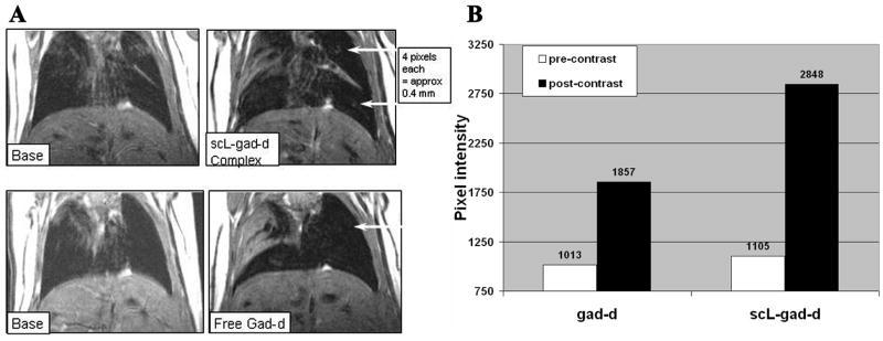
Figure 2. Detection of 4 pixel RenCa nodules.
A- Pixel Intensity of upper nodule with free gad-d was 1857, while that with scL-gad-d complex was 2848. This upper nodule does not show on base (pre-contrast image) with free gad-d and either does not show or is hidden in atelectasis on base for complex. The lower nodule is not visible except with complex. Its intensity is 2938. The region of the nodule measures 1121 with free gad-d, but the nodule itself cannot be identified. B-This chart demonstrates the increase in peak pixel intensity in the region including the small (approximately 4 pixel—400 micron) upper lung nodule shown in A

