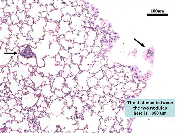
Figure 7. Histology section (H&E stain) of the lungs from the mouse described above (Figure 4A).

MR images were used to guide the sectioning of the lungs. Arrows point to the two small metastases, each less than 100 microns in size.

MR images were used to guide the sectioning of the lungs. Arrows point to the two small metastases, each less than 100 microns in size.