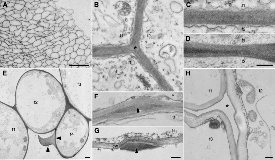
Figure 1.
Micrographs showing the existence of cotton fiber bundles. f1 to f4 indicate interiors of individual fibers, where applicable. A, Light micrograph of a cross-section of part of a fiber bundle. B to H, TEM images prepared by chemical (B, C, F, and H) or cryo (D, E, and G) fixation. During elongation, thin coherent walls (B–E) join adjacent fibers, including filling corners between three fibers (asterisk in B). E shows the distinction between the inner primary wall (arrowhead) and material between adjacent fibers (arrow) in a case where a missing fiber (from space at bottom center) likely separated from the forming bundle during specimen preparation. Occasionally, the thin joined walls are interrupted by material-filled bulges (F and G, arrows). In G, the lower fiber is missing, and the bulge material is clearly distinct from the traditional primary cell wall (cw) of the upper fiber. H shows a cleared space (asterisk) between three fibers at the onset of secondary wall thickening after the interfacial material had broken down (see below). Ages of fiber (DPA) are as follows: A, 15; B, C, and G, 10; D and E, 5; F, 20; and H, 25. Bars = 50 μm (A), 300 nm (D, for C and D; G, for F and G; and H, for B and H), and 1 μm (E).

