Figure 3.
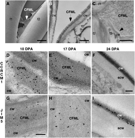
Evidence from cryo-FE-SEM and TEM analyses that the CFML is a special cell wall layer that breaks down in transition-stage fiber. Individual fibers are indicated in A to C by f1 to f3. The traditional primary cell wall of an individual fiber is indicated by cw, and the secondary wall at 24 DPA is indicated by scw. TEM immunolabeling was performed at 10 DPA (D and G), 17 DPA (E and H), and 24 DPA (F and I). In contrast to other panels including parts of two or three fibers, only one fiber is shown at 24 DPA in F and I, because CMFL degradation occurring previously led to spaces between fibers. Antibodies used were CCRC-M1 (D–F) and JIM5 (G–I). CCRC-M1 recognizes fucosylated side chains of XG, and JIM5 recognizes HG with a relatively low degree of methylesterification. A, Cryo-FE-SEM at 4 DPA showed a torn sheet of CFML material (arrow) between the inner primary walls of two adjacent fibers at a separation point. B, Similarly, TEM after cryo fixation at 10 DPA showed a distinct fringed layer (arrow) as the outer layer of the primary wall. Probably, this fiber separated from the one on the left during specimen preparation, with CFML material remaining attached to only one fiber. C, At 20 DPA during the transition, CFML polymers dispersed between fibers before they disappeared entirely (compare with Fig. 1H). The arrowhead shows a sharp separation zone between CFML polymers and the inner primary cell wall. D to F, CCRC-M1 labeled the CFML at 10 and 17 DPA, but labeling was undetectable by 24 DPA. In E, a gap was present between CFML in a bulged area and the primary cell wall of the adjacent fiber (top right corner), again indicating a distinction between these primary wall layers. G to I, JIM5 labeled the CFML at 10 and 17 DPA and the inner primary wall more lightly. However, at 24 DPA during secondary wall deposition, labeling on the primary wall that remained after CFML degradation was more intense than at earlier DPA. Bars = 5 μm (A), 200 nm (B), 1 μm (C), 300 nm (F, for D–F and I), and 150 nm (G, for G and H).

