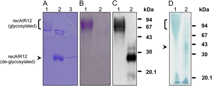
Figure 5.
SDS-PAGE (A and B), western blotting (C), and LDS-PAGE of purified recAIR12 expressed in P. pastoris. Lane 1 (A–D), Purified recAIR12 eluted from the last gel filtration chromatography purification step (9 μg for A and B, 140 ng for C, and 9 μg for D); lane 2 (A–C), same as for lane 1, but after incubation for 1 h at 37°C with EndoH; in C, recAIR12 was 47 ng; lane 3 (A), EndoH (1 μg). Gels were stained by Coomassie Brilliant Blue (A) or glycoprotein staining (B; Leach et al., 1980). C, Western-blot analysis was performed using rabbit antisera against recAIR12 expressed in E. coli (see “Materials and Methods” for details). D, RecAIR12 was treated with EndoH overnight at 22°C prior to loading on the gel (lane 2). Gel was heme stained (Thomas et al., 1976).

