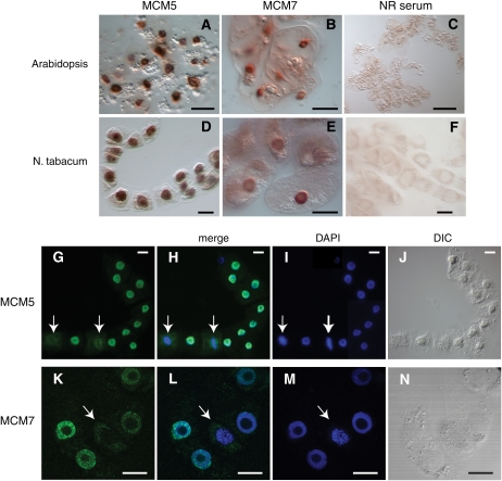
Figure 2.
Localization of endogenous MCM5 and MCM7 proteins in cultured plant cells. A to F, Immunoperoxidase staining demonstrated that MCM5 (A and D) and MCM7 (B and E) displayed localization patterns consistent with nuclear compartmentalization in both Arabidopsis and tobacco cultured cells. The discrete staining patterns for MCM5 and MCM7 were distinct from the diffuse background staining obtained using normal rabbit (NR) control serum (C and F). G to N, Immunofluorescence microscopy revealed that MCM5 (green in G and H) and MCM7 (green in K and L) colocalized with DAPI-stained DNA (blue) in most tobacco cells but not when condensed chromosomes were visible (arrows). Differential interference contrast (DIC) images are shown in J and N. The images are typical, and these patterns were observed in many cells over multiple experiments. Bars = 10 μm (A and B), 100 μm (C), and 15 μm (D–N).

