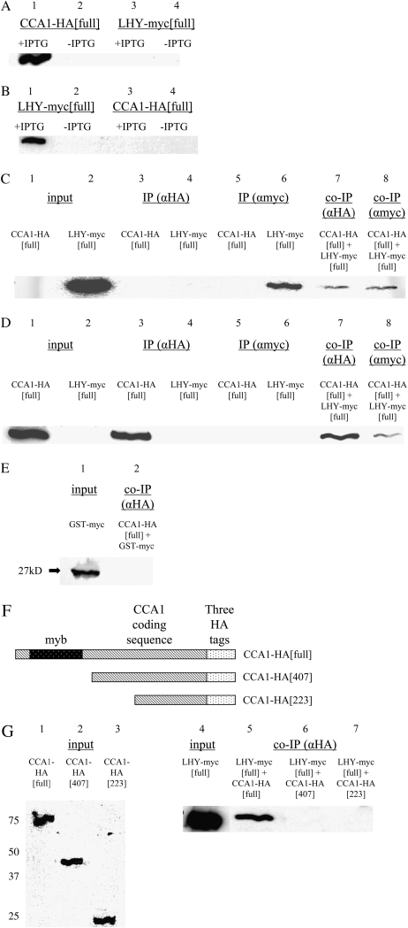
Figure 3.
CCA1 and LHY interact in vitro. A and B, E. coli transformed with CCA1-HA[full] or with LHY-myc[full] were incubated for 3 h at 37°C with or without IPTG. Proteins were extracted and equal amounts of protein were loaded into SDS-PAGE gels and analyzed by western blots with anti-HA (A) or anti-myc (B). C and D, E. coli transformed with CCA1-HA[full] or with LHY-myc[full] were incubated for 3 h at 37°C with IPTG. Extracts were immunoprecipitated with anti-HA (lanes 3 and 4) or anti-myc (lanes 5 and 6), or extracts were mixed and then coimmunoprecipitated with anti-HA (lane 7) or anti-myc (lane 8). Proteins were analyzed by western blots with anti-myc (C) or anti-HA (D). E, Protein extracts from cells of E. coli transformed with CCA1-HA[full] and cells of E. coli transformed with GST-myc were mixed and used in co-IPs with anti-HA. The proteins pulled down were analyzed on western blots with anti-myc. F, CCA1-HA[full], CCA1-HA[407], and CCA1-HA[223] constructs. G, Protein extracts from cells of E. coli transformed with CCA1-HA[full] (lane 5), CCA1-HA[407] (lane 6), or with CCA1-HA[223] (lane 7) and cells of E. coli transformed with LHY-myc[full] were mixed and coimmunoprecipitated with anti-HA. Input extract of CCA1-HA[full], CCA1-HA[407], and CCA1-HA[223] constructs analyzed by western blots using anti-HA antibodies (lanes 1–3). Input extract of LHY-myc[full] construct analyzed by western blots using anti-myc antibodies (lane 4). Co-IPs were analyzed by western blots using anti-myc antibodies (lanes 5–7).

