Figure 5.
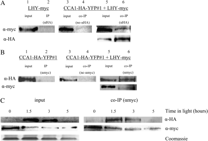
CCA1 and LHY interact in vivo. A, LHYpro∷LHY-myc LHY-null;CCA1pro∷CCA1-HA-YFP cca1-1#1 and LHYpro∷LHY-myc LHY-null plants were grown in LD for 4 weeks. Tissue was harvested and used for co-IPs with anti-HA (αHA). As a control, the co-IP experiments were also carried out without anti-HA antibody (no αHA). Proteins were analyzed by western blots with anti-myc (top row) or anti-HA (bottom row). B, LHYpro∷LHY-myc LHY-null;CCA1pro∷CCA1-HA-YFP cca1-1#1 and CCA1pro∷CCA1-HA-YFP cca1-1#1 plants were grown in LD for 4 weeks. Tissue was harvested and used for co-IPs with anti-myc (αmyc). As a control, the co-IP experiments were also carried out without anti-myc antibody (no αmyc). Proteins were analyzed by western blots with anti-HA (top row) or anti-myc (bottom row). C, LHYpro∷LHY-myc LHY-null;CCA1pro∷CCA1-HA-YFP cca1-1#1 plants were grown in LD for 2 weeks. Tissue was harvested at intervals and used in a co-IP experiment with anti-myc. The proteins were analyzed by western blots with anti-HA (top row) or anti-myc (bottom row). Input, Levels of CCA1-HA-YFP and LHY-myc in extracts. Co-IP (αmyc), CCA1-HA-YFP and LHY-myc pulled down by anti-myc antibodies. The Coomassie-stained loading control is shown below.

