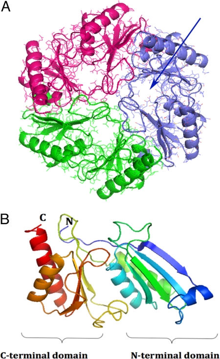
Fig. 1.
Overall structure of EutL. (A) Top view of the EutL trimer. Shown is a ribbon diagram superimposed onto an all-atom representation. The structure of each monomer is colored in blue, red, and green. The pseudo 6-fold symmetry is readily apparent as are the 3 pores in each of the monomers (blue arrow). (B) Ribbon diagram of the EutL monomer. The structure is rainbow-colored starting with the N terminus in blue and ending with the C terminus in red. The similar folds of the N-terminal and the C-terminal domains are apparent. The structure consists of residues 2–216, which could be built reliably into the electron density. The N-terminal methionine, the last 2 residues, and the His tag could not be modeled. Figs. 1–5 were generated with the program PyMol (http://pymol.sourceforge.net).

