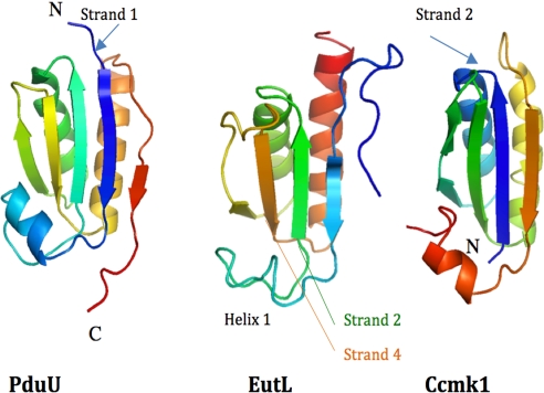
Fig. 2.
Ribbon diagram of the structures of PduU [PDB ID code 3CGI (only residues 16–122 are shown for clarity)], EutL (C-terminal domain between residues 110 and 216), and Ccmk1 (PDB ID code 3BN4). The structures are displayed in rainbow coloring starting from the N terminus in blue in each case. Helix 1 of EutL is less regularly folded and consists of only 1 helix turn. Even though the overall fold of all proteins is very similar, the order of secondary structure elements is different. Whereas the structures EutL and PduU begin with and N-terminal β-strand (arrows), in the structure of Ccmk1 this strand and former helix 1 are now positioned at the C terminus.

