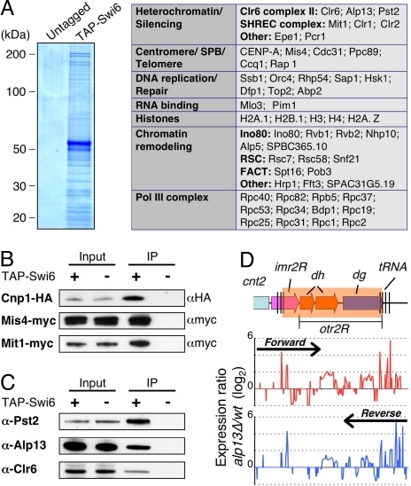
Fig. 2.
Swi6 associates with the Clr6 HDAC, and factors involved in chromosome segregation. (A) Coomassie blue staining of TAP Swi6 from cells expressing amino-terminally TAP-tagged Swi6 and untagged Swi6 are shown. The purified proteins were subjected to tandem MS (LC-MS/MS) analyses. The identified nuclear proteins were sorted into functional groups, as indicated in the table. (B and C) Fractions immunoprecipitated from indicated strains were subjected to Western blot analyses by using α-HA antibody (12CA5) to detect Cnp1-HA, α-myc antibody (9E10) to detect Mis4-myc or Mit1-myc (B) or α-Alp13, and α-PstII and α-Clr6 antibodies (C) to detect Clr6 complex II. (D) Expression profiling at the right pericentromeric repeat region of cen2 was performed for the wild-type and alp13Δ cells. Expression ratios (mutant/wt) for forward strand (Upper, red) and reverse strand (Lower, blue) probes were plotted on a log2 scale.

