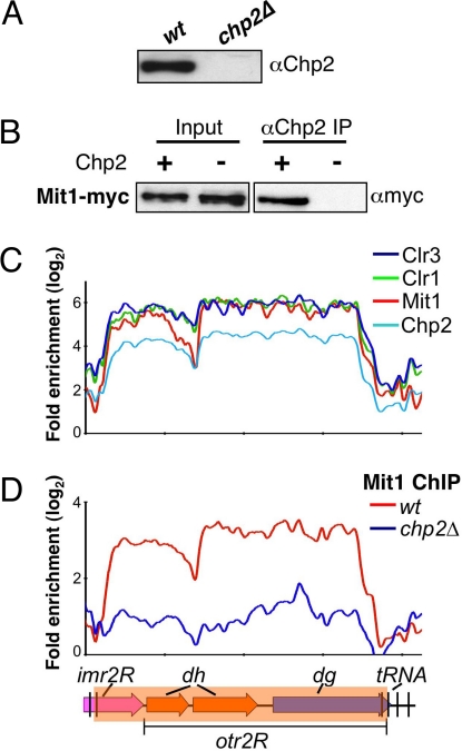
Fig. 3.
Chp2 interacts with SHREC. (A) The antibody specifically recognizes Chp2 protein. Extracts prepared from wt and chp2Δ strains were analyzed by Western blot analysis using α-Chp2 antibody. (B) Mit1, a component of the SHREC interacts with Chp2. Extracts prepared from wt and chp2Δ cells expressing Myc-tagged Mit1 were immunoprecipitated using the α-Chp2 antibody. Immunoprecipitated (IP) fractions were analyzed by Western blot analysis using α-myc antibody. (C) Chp2 colocalizes with SHREC subunits. Distribution profiles of indicated factors were determined by ChIP-chip. (D) Chp2 dependent localization of Mit1 at centromeric repeats. Mit1-myc distributions in wt or in chp2Δ backgrounds are shown. The probes are aligned to correspond with the schematic representation below.

