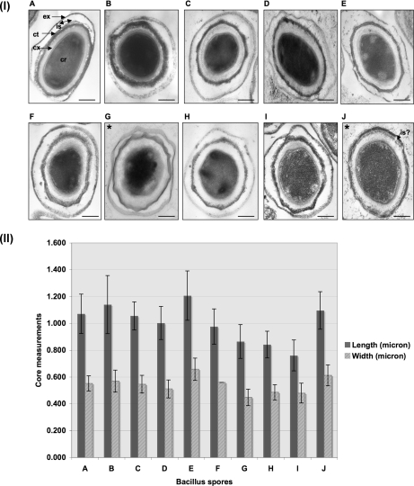
Fig. 3.
Panel I, TEM showing ultrastructural analysis of B. anthracis and closely related strains. A, B. anthracis Sterne 34F2; B, B. thuringiensis BGSC 4AJ1; C, B. cereus m1293; D, B. cereus BGSC 6E1; E, B. cereus ATCC 10987; F, B. cereus ATCC 10876; G, B. thuringiensis BGSC 4Y1; H, B. cereus m1550; I, B. mycoides DSMZ 2048; J, B. pseudomycoides DSMZ 12442. ex, exosporium; is, interspace; ct, coat; cx, cortex; cr, core. The bar is 100 nm at magnification ×60,000. All the spores were prepared using the modified G medium at 37 °C up to 72 h at 250 rpm and purified using a 50% renograffin gradient. Panel II, core dimensions of the spores of closely related Bacillus strains. A–J show the same strains listed for panel I. Error bars represent standard deviation in microns.

