
Fig. 5.
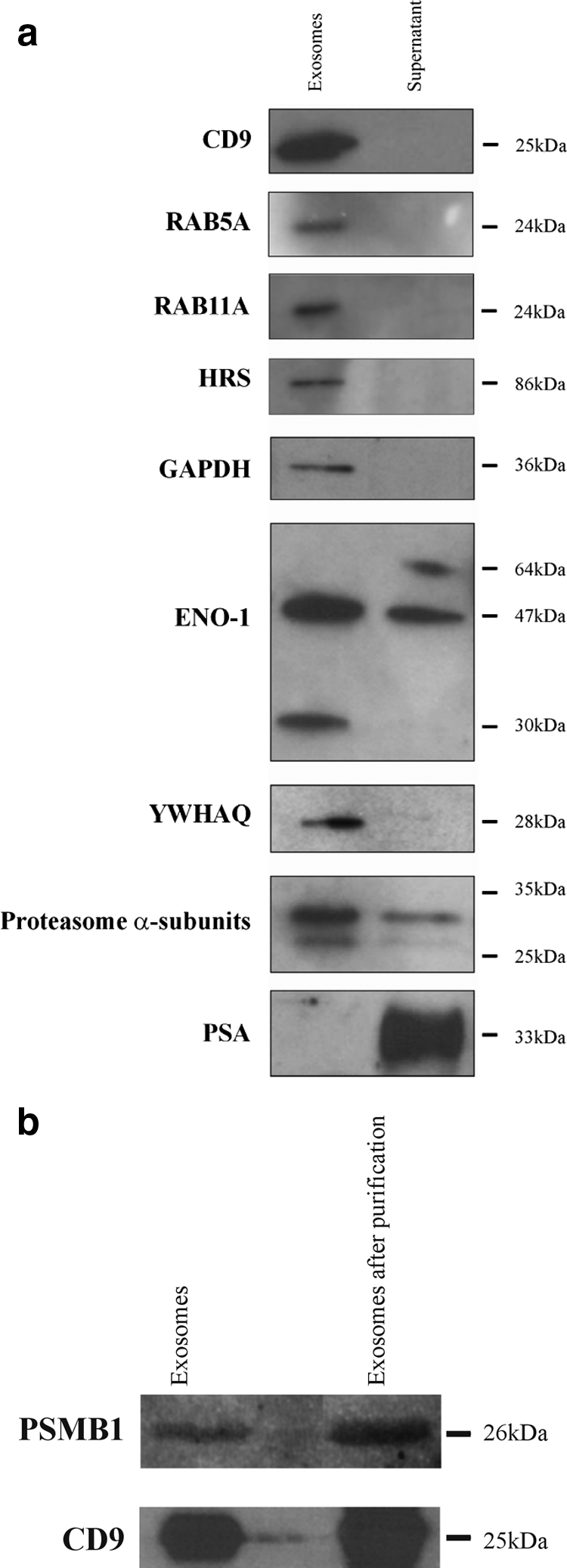
a, 1D PAGE and Western blotting analysis comparing the exosome and supernatant fractions of the PC346C cell line for CD9, the members of the RAS oncogene family RAB5A and RAB11A, HRS, GAPDH, ENO1, YWHAQ (14-3-3 protein θ), proteasome α subunits, and PSA. CD9, RAB5A, RAB11A, HRS, GAPDH, and YWHAQ were uniquely identified in the isolated exosomes, whereas PSA could only be detected in the supernatant of the PC346C cell line. b, 1D PAGE and Western blotting analysis of the exosome fraction after purification with magnetic beads. This figure shows that the CD9 and proteasome β1 (PSMB1) signals are visible in the exosome fraction both before and after purification with magnetic beads, indicating that proteasome subunits are present inside exosomes or exosomal membranes.