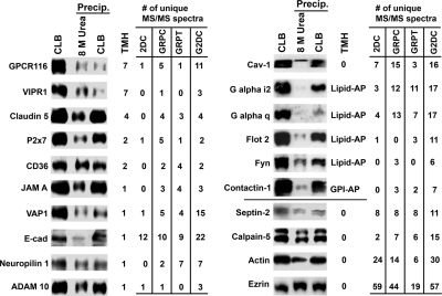
Fig. 7.
Protein partitioning during sample processing for 2DC. An immunoblot of plasma membrane proteins treated with different procedures and reagents is shown. Membrane samples were solubilized directly in CLB for SDS-PAGE, or CLB-solubilized proteins were first precipitated and then resolubilized by either 8 m urea as for 2DC or CLB. The resolubilized samples from the precipitations (Precip.) were centrifuged, and the supernatants were collected for SDS-PAGE. The number of putative or known transmembrane helices (TMH) and number of unique peptides detected by each method are shown in the table on the right. 0*, partially membrane-embedded with high hydrophobic region; Lipid-AP, lipid-anchored proteins; GPI-AP, glycosylphosphatidylinositol-anchored proteins. See supplemental Table 1 for information on antibodies and protein names. VE-cad, VE-cadherin; JAM A, junctional molecule A.

