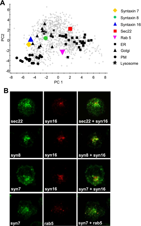
Fig. 5.
Intracellular distributions of unassigned proteins. A, the positions of several proteins of interest were marked on a PCA plot of the complete dataset: sec22, red square; syntaxin 7, yellow diamond; syntaxin 8, green circle; rab 5, magenta inverted triangle; syntaxin 16 (a Golgi marker protein), blue triangle. Black shapes indicate the marker proteins of the plasma membrane (dots), Golgi (triangles), lysosome (stars), and ER (squares). B, immunofluorescence microscopy was used to compare the distribution of sec22, syntaxin 8, and syntaxin 7 with the Golgi marker syntaxin 16. Syntaxin 7 distribution was also compared with that of the early endosome marker Rab 5.

