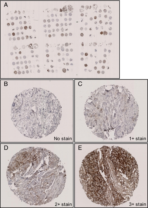
Fig. 7.
Immunohistochemical staining of EMMPRIN. EMMPRIN immunohistochemical staining was performed on an independent sample set of 156 tissues using TMA. A, overview of TMA; B, negatively stained tissue; C, 1+ membrane stain; D, 2+ stain; E, 3+ stain. Overview picture was taken at 5× magnification; other pictures were taken at 100× magnification.

