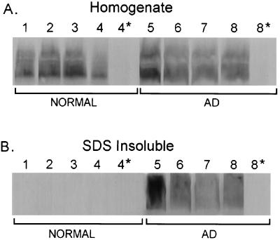
Figure 2.
Western blot analysis of agrin expression in normal aged and AD) brain. Equal volumes of total homogenates or of the SDS-insoluble fractions from normal (1–4) or AD brain (5–8) prefrontal cortex (area A10) were probed with anti-agrin antisera. All samples were solubilized in 0.2 M NaOH before electrophoresis (see Materials and Methods). (A) The anti-agrin antibody recognized a polypeptide with an apparent mobility of ≈500 kDa in both normal and AD brains. No polypeptide was detected when normal rabbit IgG was substituted for the first layer (lanes 4* and 8*). (B) Virtually all of the agrin from normal brains was efficiently solubilized in 1% SDS at neutral pH (lanes 1–4). However, a portion (see Results) of the agrin from AD brains was insoluble under these conditions (lanes 5–8). No polypeptide was detected when normal rabbit IgG was substituted for the first layer (lanes 4* and 8*).

