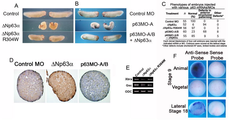
Figure 2. ΔNp63 loss and gain of function results in anterior patterning defects in Xenopus laevis.
(A) Four-cell Xenopus embryos were injected dorsally with ΔNp63 α or ΔNp63 -R304W mRNA (2 ng each) and scored at the tailbud stage. (B) Four-cell Xenopus embryos were injected dorsally with p63MO-A (200 ng) in the presence or absence of ΔNp63 α mRNA (2 ng), and scored at the tailbud stage. (C) Table summarizing results from embryo injections in (A) and (B). (D) Animal caps injected with control or p63MO, or ΔNp63-encoding RNA were fixed, sectioned, and stained for p63 (4A4 antibody). (E) Whole embryo RT-PCR shows that the pan-mesodermal marker Xbra and the anterior mesodermal marker Gsc are down-regulated in ΔNp63 α-injected but not ΔNp63 α-R304W-injected embryos. (F) In situ hybridization analysis shows that ΔNp63 is expressed in the animal pole of late blastula (stage 9) embryos and in most of the epidermis of mid-neurula (stage 18) embryos (anterior is on the left).

