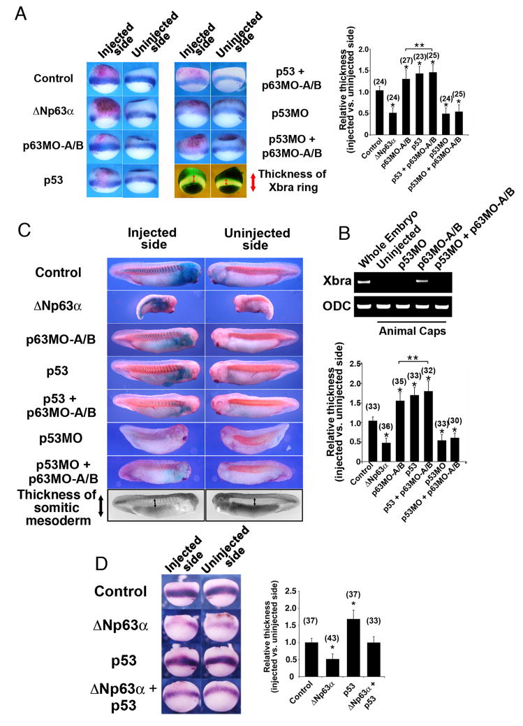
Figure 4. Mesoderm induction due to loss of p63 is p53-dependent.
(A) Embryos were injected with ΔNp63 mRNA (2 ng), p63MO-A/B (200 ng), p53 mRNA (100 pg), p53MO (30 ng), p53 mRNA + p63MO-A/B, or p53MO + p63MO-A/B. The relative extent of the Xbra expression domain between the injected and uninjected sides of each embryo was quantified as shown (two lower right image panels) and plotted on a bar graph. β-galactosidase mRNA (β-gal, 250 pg) was co-injected to mark the injection site. (B) RT-PCR analysis in animal caps injected with p63MO, p53MO, or both morpholinos confirms that down-regulation of p63 can induce mesoderm (as assessed by the pan-mesodermal marker Xbra) only in the presence of p53. (C) Embryos were injected as in (A), fixed at stage 26, stained for β-galactosidase activity (X-gal, blue color), and processed for whole-mount immunohistochemistry with an antibody specific for somitic mesoderm (12/101). The relative extent of the somitic mesoderm between the injected and uninjected sides of each embryo was measured as shown (two lower right image panels) and plotted on a bar graph. (D) Embryos were injected with ΔNp63 mRNA (2 ng), p53 mRNA (100 pg), or both, and β-galactosidase mRNA (β-gal, 250 pg) was co-injected to mark the injection site. The relative extent of the Xbra expression domain between the injected and uninjected sides of each embryo was quantified as in (A) and plotted on a bar graph. For (A) and (D), injected embryos were fixed at stage 10.5, stained for β-galactosidase activity (Red-gal, red color) and processed for in situ hybridization with probes for the pan-mesodermal marker brachyury (Xbra). Analysis of the extent of mesoderm tissue formation for (A), (C), and (D) is described in the methods section. *, **: single asterisks indicate significant changes from control embryos; double asterisks indicate significant changes between p63 MO and p63MO + p53 injected embryos (Student’s t-test, p<0.05). Numbers in parentheses indicate numbers of embryos scored.

