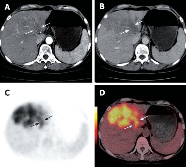
Figure 3.

Contrast CT scan demonstrating a left lobe mass and portal vein thrombus in a 60-year-old woman. During the arterial phase, the thrombus appears as a hypodense intraluminal area with dense linear enhancement (white arrows, A). A filling defect was detected in the left branch during the portal phases (white arrows, B). PET (black arrows, C) and PET/CT fused images (white arrows, D) reveal a highly metabolic thrombus in the left branch.
