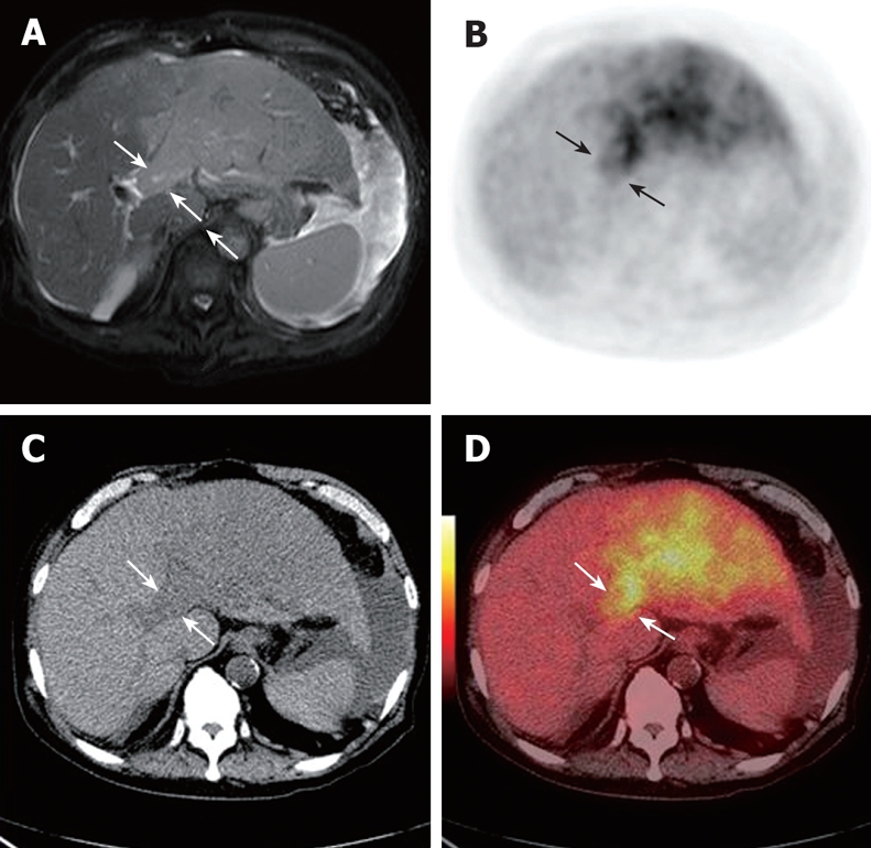
Figure 4.

MRI scan showing a left lobe mass and portal vein thrombus in an 80-year-old man (white arrows, A). CT detected a low-density lesion in the left branch (white arrows, C). PET (black arrows, B) and PET/CT fused images (white arrows, D) reveal a highly metabolic thrombus in the left branch.
