
Figure 2.
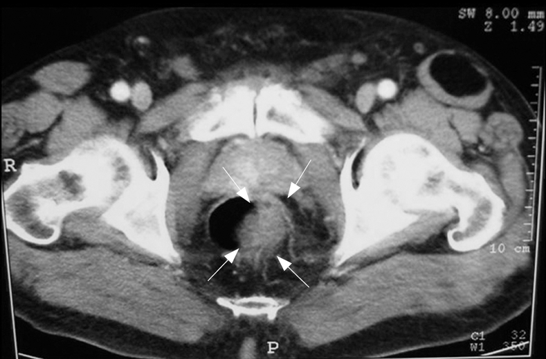
TC confirming the sonographic findings of the presence of a mass with a marked, irregular, eccentric thickening of the lateral left wall of the lower third of the rectum, but providing no evidence for either pelvic lymphadenopathy or distant metastasis.