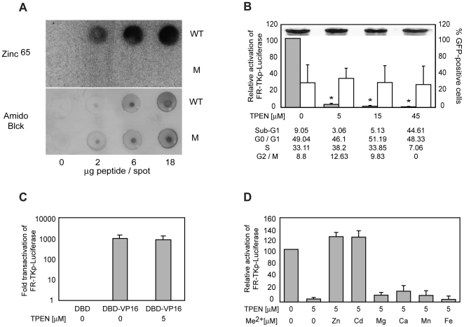
Figure 2. Zinc is coordinated by EBNA1's UR1 domain, and is required for EBNA1 to transactivate.
(A) Evaluation of Zinc65 binding by the wild type (WT) and mutant (M) UR1 peptides. The indicated amounts of WT and M peptides were dot-blotted on a 0.2 mM PVDF membrane, and probed with radioactive zinc (∼50 µCi) as described in the Methods section. Washed membranes were dried and visualized using a Phosphorimager. WT peptide was observed to bind Zn65 in a concentration-dependent manner. In contrast, the M peptide failed to bind zinc. To confirm that both peptides bound the PVDF membrane, a blot prepared in parallel was dried and stained using Amido Black. (B) The metal chelator TPEN reduces the ability of EBNA1 to transactivate FR-TKp-Luciferase. C33a cells were co-transfected the FR-TKp-Luciferase reporter plasmid, an EBNA1-expression plasmid, and a CMV-EGFP expression plasmid. Cells were treated with the indicated levels of TPEN at the time of transfection, and harvested 15 hours later. Harvested cells were analyzed by flow cytometry to determine the fraction of live-transfected cells, the level of EGFP expression in that fraction. For cell-cycle analysis, the cells were fixed and then PI-stained. An aliquot of harvested cells were used to determine the expression level EBNA1 by immunoblot upon treatment with the indicated amounts of TPEN, shown as an inset in the graph. The rest of the cells were processed to determine luciferase activity, which is expressed as a percent of the luciferase activity observed in the absence of TPEN treatment, and shown in the grey bars. The open bars indicate the percent of live EGFP-positive cells at each concentration of TPEN. The asterisks indicate that the significantly lower levels of luciferase activity were observed in the presence of 5, 15 and 45 µM TPEN (p<0.05, Wilcoxon rank-sum test). The cell-cycle profile of TPEN-treated and control cells was obtained for one experiment, and is shown below the graph. (C) TPEN does not affect transactivation by DBD-VP16. C33a cells were co-transfected with the FR-TKp-Luciferase reporter plasmid, and expression plasmids for DBD, and DBD-VP16. Some DBD-VP16 transfected cells were treated with the indicated amounts of TPEN at the time of transfection, and analyzed 15 hours later to determine the fraction of live-transfected cells, and the level of luciferase expression. The activity observed with DBD-VP16 in the absence and presence of TPEN is expressed as the fold-over the luciferase level observed with DBD alone. (D) Zinc and Cadmium reverse TPEN-mediation inhibition of transactivation by EBNA1. C33a cells were co-transfected with the FR-TKp-Luciferase reporter plasmid, an EBNA1 expression plasmid, and a CMV-EGFP expression plasmid, and treated with 5 µM TPEN at the time of transfection. Fifteen hours post-TPEN addition, 5 µM of Zn (CH3COO)2, Cd(CH3COO)2, CaCl2, MgSO4, MnCl2, or Fe(CH3COO)2, was added to the cells for an additional 15 hours. Cells were harvested and analyzed by flow cytometry to determine the level of live-transfected cells, followed by determination of luciferase activity. The relative activation is shown in the grey bars, and is expressed as a percent of the luciferase activity observed in the untreated sample.

