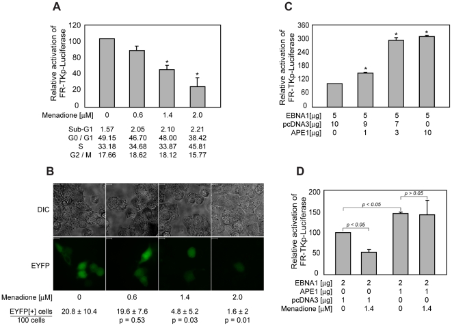
Figure 6. Transactivation by EBNA1 is sensitive to oxidative stress, and is augmented by over-expression of APE1/Ref-1.
(A) Menadione reduces the ability of EBNA1 to transactivate FR-TKp-Luciferase. C33a cells were co-transfected the FR-TKp-Luciferase reporter plasmid, and an EBNA1-expression plasmid. Cells were treated with the indicated levels of menadione six hours post-transfection, and harvested 18 hours later. For cell-cycle analysis, an aliquot of cells was fixed and then PI-stained. The rest of the cells were processed to determine luciferase activity, which is expressed as a percent of the luciferase activity observed in the absence of menadione treatment, and shown in the grey bars. The cell-cycle profiles of menadione-treated and control cells were obtained for one experiment, and are shown below the graph. The asterisks indicate statistical significance (p<0.05) compared to control. (B) Menadione affects the ability of UR1 to support bimolecular fluorescence complementation. C33a cells were transfected with expression plasmids for A) EYFP(2-154) and EYFP(155-238), or B) & C) expression plasmids for UR1-EYFP(2-154) and UR1(155-238). Transfected cells were treated with vehicle alone, or 0.6, 1.4 and 2.0 µM menadione six hours after transfection. Cells were visualized by fluorescence for EYFP (YFP) or by light microscopy (DIC) after 18 hours of menadione treatment. The scale bars indicate a length of 10 microns. The fraction of fluorescent cells observed for each menadione concentration in five independent measurements is shown along with the standard deviation. The p-values indicate statistical significances calculated using the Wilcoxon rank-sum test upon a pair-wise comparison to untreated cells. p-values lower than 0.05 indicate significance. (C) Over-expression of Ref-1/APE1 augments transactivation by EBNA1. C33a cells were transfected with an EBNA1 expression plasmid, and increasing amounts of an expression plasmid for Ref-1/APE1 along with the FR-TKp-luciferase reporter plasmid. Luciferase levels were evaluated 48 hours post-transfection, and are expressed as a fraction of transactivation observed in the absence of Ref-1/APE1, which was set to be 100%. The asterisks indicate statistical significance (p<0.05) compared to control. (D) Over-expression of Ref-1/APE1 ameliorates the effect of menadione on EBNA1 mediated transactivation. C33a cells were transfected with an EBNA1 expression plasmid, and either 1 µg of a Ref-1/APE1 expression plasmid or empty vector control plasmid in addition to the FR-TKp-luciferase reporter plasmid. Six hours post-transfection, the cells were split and half the cells were treated with 1.4 µM of menadione. Luciferase levels were evaluated 24 hours post-transfection. p values are indicated in the figure.

