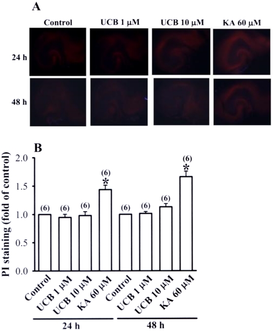
Figure 6. Effects of prolonged UCB exposure on propidium iodide (PI) staining in hippocampal slice cultures.
(A) Representative images of PI staining in slices from control or treatment with UCB (1 µM or 10 µM) or kainic acid (60 µM) for 24 or 48 h. (B) Densitometry quantification of PI staining similar to those shown in (A). Data are expressed as fold of increase over the respective control group. Number of experiments is indicated in the parenthesis.

