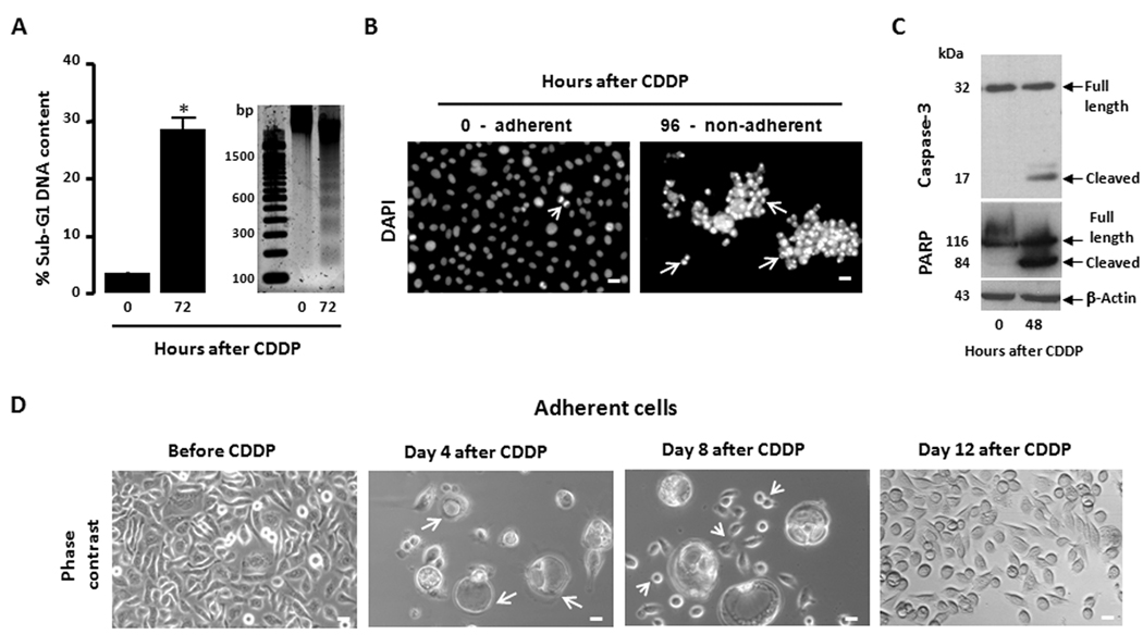
Figure 1.
Lethality of 1-h exposure of OV2008 cells to 20 µM CDDP and the fate of residual cells that survive treatment. OV2008 cells were seeded and allowed to attach to the plate surface for 48 h. Cells were exposed to either vehicle (0.9 % NaCl) or 20 µM CDDP for 1 h and cultured in CDDP-free media thereafter. (a) 72 h following CDDP exposure, the cells were pelleted, and either permeabilized, labeled with propidium iodide, and the sub-G1 DNA content assessed by microcapillary cytometry, or the genomic DNA was isolated, separated by agarose electrophoresis, stained, and photographed. Bar, mean ± SEM. *, P < 0.05 relative to values at 0 h. (b) 96 h after CDDP treatment, the non-adherent cells were re-adhered to a microscope slide, stained with DAPI, photographed using a fluorescence microscope and compared against adherent cells of vehicle-treated cultures. Arrows, apoptotic nuclei. Arrowhead, cell undergoing mitosis. Scale bar = 20 µm. (c) 48 h after CDDP exposure, whole protein extracts where obtained and separated by electrophoresis, and immunoblots were probed with the indicated antibodies. (d) Cells that remained adherent following exposure to CDDP are shown as photographed by phase contrast microscopy. Arrows, large vacuolated cells likely undergoing degeneration. Arrowheads, cells undergoing repopulation. Scale bar = 20 µm.

