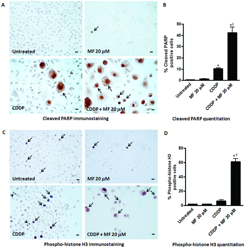
Figure 6.

Effect of a cytostatic concentration of MF on the expression of cleaved poly (ADP-ribose) polymerase (PARP) and phospho-histone H3 in OV2008 cells studied respectively 8 or 10 days after being exposed to a lethal concentration of CDDP for 1 h. Cells were seeded for 48 h, and then treated with vehicle (0.9 % NaCl) or 20 µM CDDP for 1 h. CDDP was then removed and fresh media without or with 20 µM MF was replaced every 2 days. As control groups, untreated cells in their logarithmic phase of growth, and cells that received only 20 µM MF for 8 or 10 days were included in the study. At the end of the experiment, adherent cells were washed, fixed with 4% paraformaldehyde, and subjected to immunocytochemistry using an antibody that recognizes only the cleaved form of PARP (a), or phospho-histone H3 (c). Quantitation of cleaved PARP (b) and phospho-histone H3 (d) positive nuclei were done using a microscope with 400 X magnification and at least 500 cells were counted for each sample. The experiment was done in triplicates and the results are expressed as percentage of cleaved PARP or phospho-histone H3 positive cells. Arrows indicate examples of positive labeling. In panel (a) notice that in cultures receiving CDDP + MF 20 µM, cleaved PARP positive staining is observed not only in large degenerated cells, but also in smaller cells. In panel (c), arrowheads represent figures of mitosis. Bars, mean ± SEM. Scale bar = 20 µm. *, P < 0.05 compared to untreated; †, P < 0.05 compared to CDDP.
