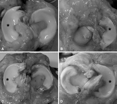
Fig. 1A–D.
Photographs show menisci at 4 months in (A) contralateral control, (B) meniscectomized, (C) aseptic allograft, and (D) sterile allograft. A synovial-derived connective tissue covers the periphery of the plateau in the meniscectomized joint. Both allografts show laxity in their attachment and displacement toward the periphery. *Lateral meniscus.

