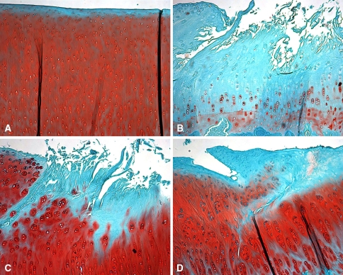
Fig. 5A–D.
Histologic features of the tibial plateau are shown in these photomicrographs of sections of tibia from (A) control, (B) meniscectomized, (C) aseptic allografted, and (D) sterile allografted knees that were stained with safranin O for proteoglycans (original magnification, ×40). Surgically treated knees had fissuring and fibrillation of the surface accompanied by a decrease in proteoglycan staining. Allografted knees had chondrocyte cloning with intense proteoglycan staining in regions adjacent to damage.

