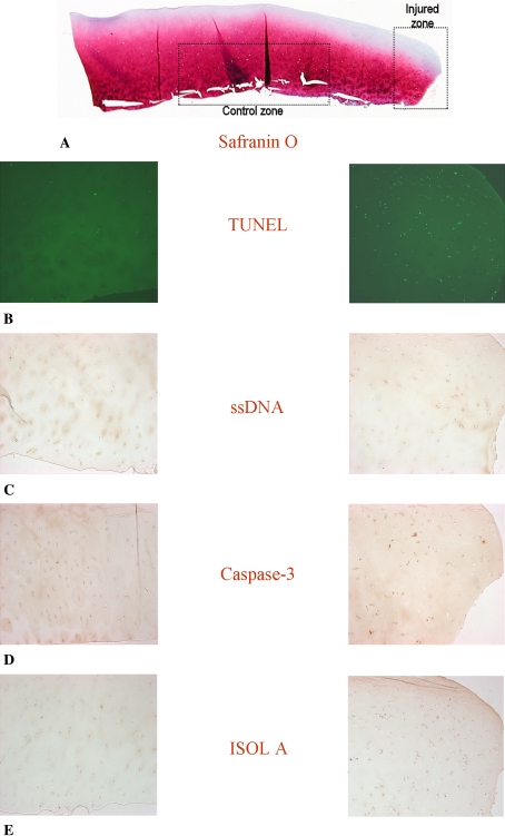
Fig. 1A–E.
Photomicrographs (Stain, Safranin O (A) and DAB primary antibody staining with Fast Green counter stain (B–E); original magnification, ×10) compare (A) the injured zone and the control zone in (B) TUNEL, (C) ssDNA, (D) anti-active caspase-3, and (E) ISOL. The break in the cartilage is to the right of the injured zone images. TUNEL = terminal deoxynucleotidyl transferase end labeling; ssDNA = DNA denaturation analysis using anti single-stranded DNA antibody; ISOL = in situ oligonucleotide ligation.

