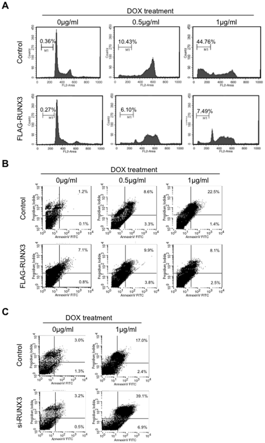
Figure 6. RUNX3 overexpression inhibited chemotherpeutic drug induced apoptosis.
A: Adriamycin (Dox, 0, 0.5 and 1 µg/ml) was treated for 72 hours in control and RUNX3 overexpressing HSC3 cells. Cell cycle distribution was determined by DNA content analysis after propidium iodide staining using a flow cytometer. For each sample, 20,000 events were stored. Percentage of sub-G1 population is indicated. We performed two independent experiments with triplicate wells per condition. Representative data is shown. B: Flow cytometric analysis of Annexin V and propidium iodide staining in control and RUNX3 overexpressing cells after DOX treatment for 72 h at the indicated doses. C: Flow cytometric analysis of Annexin V and propidium iodide staining in control and RUNX3 knockdown cells after DOX treatment for 72 h at the indicated doses. We performed two independent experiments.

