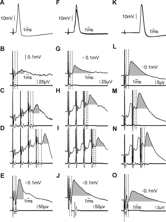
Figure 2.
Cervical spinal motoneurons receive monosynaptic and disynaptic reticulospinal input. A–E, Example motoneuron projecting to forearm flexors that received disynaptic reticulospinal contacts. A, Antidromic activation from median nerve above the elbow (overlain single sweeps). B–D, Disynaptic reticulospinal EPSPs following single 300 μA MLF stimulus (B), train of three stimuli (C), and train of four stimuli (D). Each panel shows averaged intracellular records (top) with simultaneously recorded epidural volleys below. Vertical dashed lines highlight the segmental latency of the response. Calibrations in B apply to B–D. E, EPSP evoked in this cell following single 300 μA stimulus to PT. F–J, Example monosynaptic EPSPs evoked following reticulospinal activation in a spinal motoneuron also projecting to forearm flexors. Panels and plotting conventions are as in A–E. K–O, Example motoneuron projecting to thenar muscles which received powerful monosynaptic input from the reticulospinal tract. Panels and plotting conventions are as in A–E, except that responses to a train of two and three stimuli are shown in M and N, respectively.

