
Fig. 5.
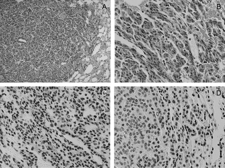
Hematoxylin and eosin and immunohistochemical staining of lung tissue from DOX/BHT-treated mice. Panels (A) and (B) are ×10 and ×40 magnifications, respectively, of hematoxylin and eosin-stained slides showing mild edema around the tumor; panels (C) and (D) are immunohistochemical staining of lung tissue slides with antibodies to COX-2 and NF-κβ (Ser276) showing COX-2 and NF-κβ positivity in blood vessels and stromal cells, respectively; magnification for IHC ×40.