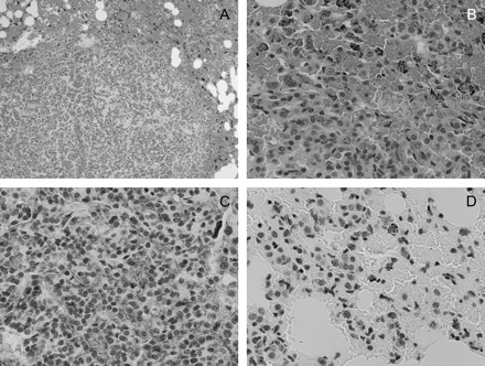
Fig. 6.

Hematoxylin and eosin and immunohistochemical staining of lung tissue from DOX/curcumin-treated mice. Panels (A) and (B) are ×10 and ×40 magnifications, respectively, of hematoxylin and eosin-stained slides showing marked inflammation around the tumors and a high density of red blood cells suggestive of edema (panel A) with infiltration of macrophages (panel B, cells with black dots in panel D). (C) Immunohistochemical staining of lung tissue slides with an antibody to COX-2. The tumor has a high-density vasculature that contains endothelial cells staining positively for COX-2. (D) Immunohistochemical staining of lung tissue slides with an antibody to NF-κβ (Ser276) demonstrating NF-κβ positive macrophages infiltrating the tumor tissue; magnification for IHC ×40.
