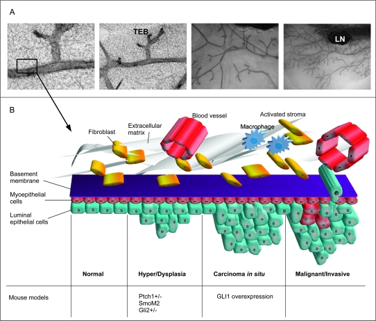
Fig. 2.
The Hh pathway and mammary gland tumour formation. (A) The structure of the normal mouse virgin mammary gland, whole mounts depicting the ductal tree and the terminal end buds are shown in increasing resolution from right to left. (B) A schematic illustration of cancer progression in the mammary gland in relation to mouse models with a de-regulated Hh pathway. Early lesions appear in the form of hyper and/or dysplasias in the mammary epithelium. These changes are seen in mice haploinsufficient for Ptch1 and in mice with targeted expression of a constitutively active form of Smo in the mammary gland (26,40). Hyper and dysplasias are also formed when normal mammary stem cells over-expressing Gli2 are transplanted into immunodeficient hosts (39). In the next step called carcinoma in situ (CIS), transformed cells appear in the dysplastic areas, but no invasive growth or metastases are observed. Such tumours are induced by targeted over-expression of GLI1 in the mammary gland (M.Fiaschi, B.Rozell, Å.Bergström, R.Toftgård, submitted). Development of fully malignant tumours induced by activated Hh signalling has not yet been reported. TEB, terminal end bud; LN, lymph node.

