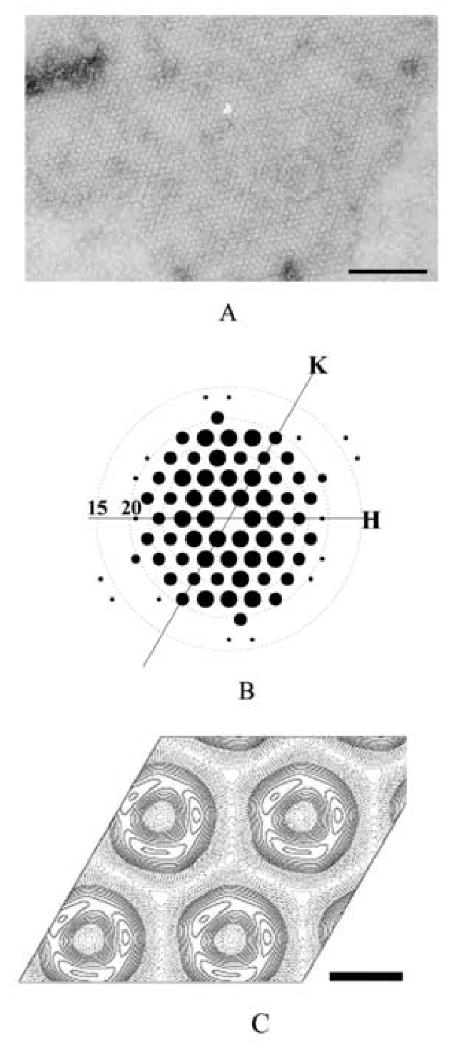
Figure 1. 2D crystals of SecA ring-like structures.

A. The negative stained 2D crystals of SecA formed on E. coli lipid monolayers. The crystallization condition was 10 mM Tris-Ac, pH 7.4, 200 μg/ml SecA, incubating with lipid monolayer composed of E. coli lipid extract for 24 hour at 4°C. The scale bar is 100 nm.
B. Computed Fourier transform of a SecA 2D crystal after two-round unbending. The resolution circles representing 20, 15 Å were indicated. Only spots with IQ less than 4 are displayed, and the size of the spot is inverse proportional to IQ: IQ 1 is largest and IQ 4 is smallest.
C. The projection map of SecA 2D crystals averaged from 10 untilted images without any symmetry imposed. The scale bar is 5 nm.
