
Figure 3.
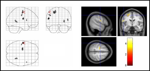
Areas of increased regional white matter volumes in TS patients compared with controls. Areas of increased regional white matter volumes in TS patients compared with controls. The same conventions apply as for figure 1. Significant voxels were found in the left primary sensorimotor cortex, and left middle frontal gyrus (corrected P < 0.05). Note that changes in the right parahippocampal gyrus, which were not significant, are also displayed. TS = Tourette syndrome.