
Figure 4.
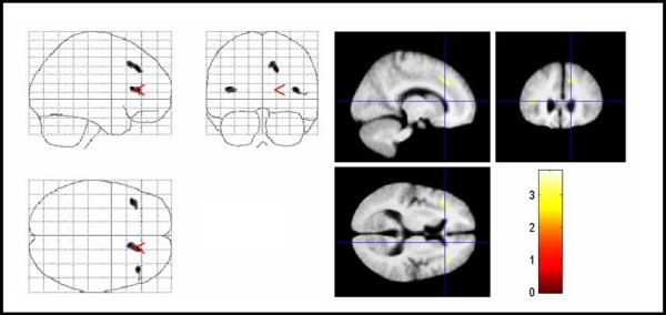
Areas of decreased MTR in TS patients compared with controls. Areas of decreased MTR in TS patients compared with controls. The significant results are superimposed on the averaged normalized MTR maps of the study population (thresholded at uncorrected P < .005 for display purpose only). Significant voxels were found in the right medial frontal gyrus, inferior frontal gyrus bilaterally, and the right cingulate gyrus (corrected P < .05). TS = Tourette syndrome, MTR = magnetization transfer ratio.