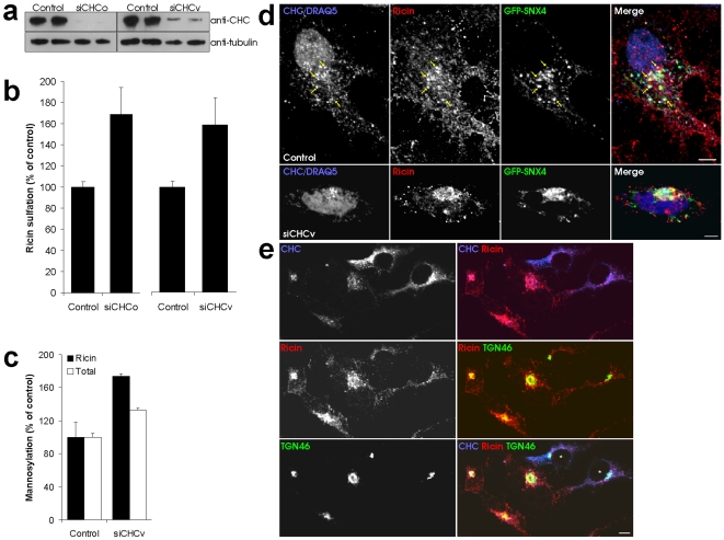
Figure 5. CHC knockdown leads to increased ricin transport.
(a) Transfected cells were lysed and analyzed by Western blot with the indicated antibodies. (b) Cells transfected as indicated were incubated with radioactive sulphate for 3 h before further incubation with ricin sulf-1 for 90 min and subsequent lysis. Ricin was immunoprecipitated from the lysates, and the precipitate separated by SDS-PAGE before autoradiography. The intensity of the bands was quantified and the average plotted with error bars showing standard deviations. (c) Transfected cells were incubated with [3H]mannose in glucose free medium for 3 h before further incubation with ricin sulf-2 for 3 h. Cell lysate was immunoprecipitated with anti-ricin antibodies. The precipitate was analyzed by autoradiography. (d) Cells transfected as indicated were incubated with 2 µg/ml ricin for 45 min before fixation and staining with antibodies as indicated. Bars, 5 µm. (e) Cells transfected with siCHCv were incubated with 2 µg/ml ricin for 45 min before fixation and staining with the indicated antibodies. Asterisks in the merged picture indicate control cells. Bar, 5 µm.

