Figure 1.
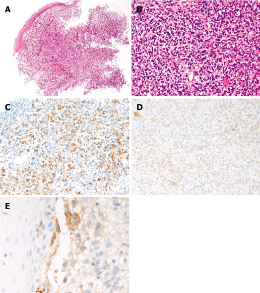
Esophageal amelanotic malignant melanoma in Case 1. A: Low power view of the biopsy (HE, × 20); B: High power view of the biopsy. Malignant polygonal and spindle cells are seen. No melanin pigment is seen (HE, × 200); C: The tumor cells are positive for melanosome (HMB45). (Immunostaining, × 200); D: The tumor cells are positive for S100 protein (Immunostaining, × 200); E: The tumor cells are focally positive for KIT (Immunostaining, × 200).

